

A clearer picture of your meniscus
Knee operations on the meniscus present challenges because its tissue is only partially supplied with blood. Researchers are creating a new three-dimensional map to reduce the risks.
Related news
Technology that moves you

The wheelchair of the future – made in Switzerland

Keeping one step ahead of the next infectious disease
The meniscus around the knee joint is a complicated tissue that is only supplied with blood in certain areas. However, precise knowledge of this valuable vascular plexus is important as blood supply is crucial to the self-healing potential of the meniscus. Until now, our understanding has relied on outdated technology, over 40 years old, that produces only two-dimensional images of tissue sections. According to press release by the Swiss Federal Laboratories for Materials Science and Technology (Empa), this means that valuable data on the deformability of the cartilage or the vascular network, for example, is lost.
3D map of a meniscus
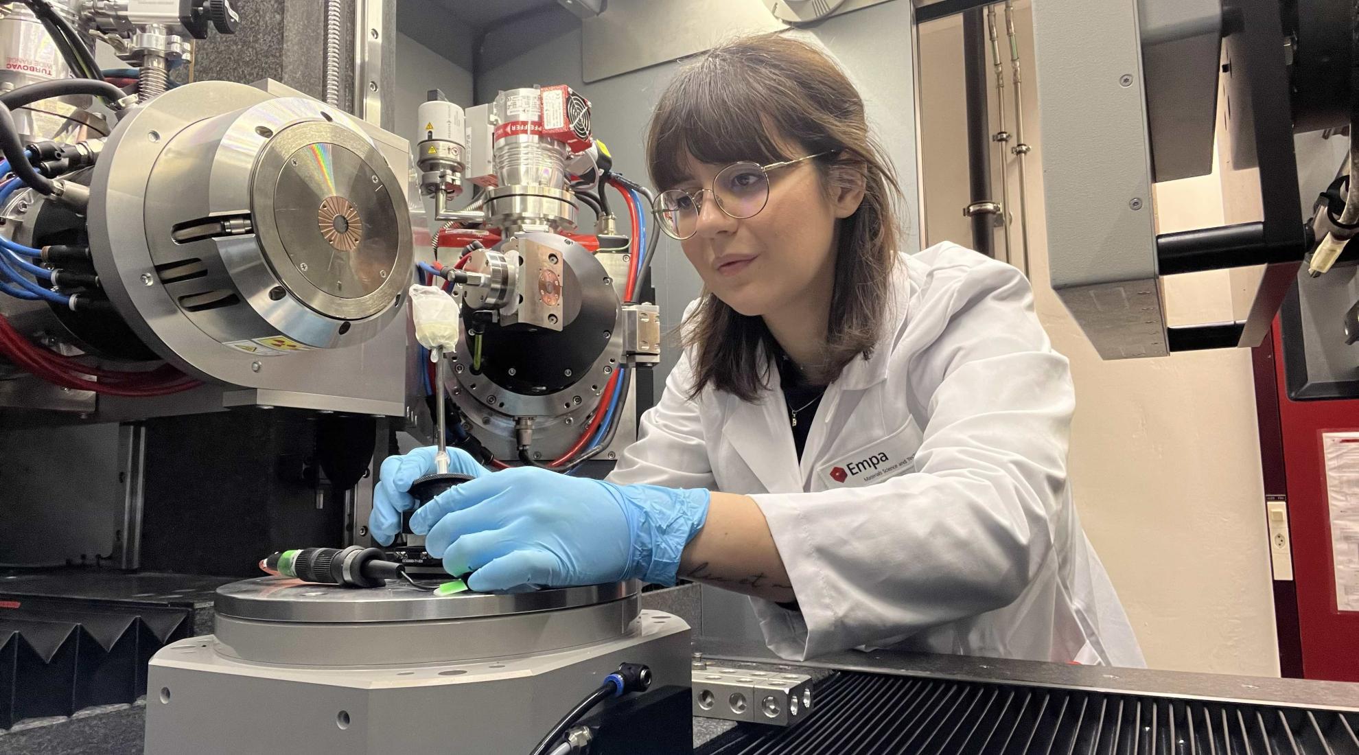
Empa researchers are developing advanced 3D technology to map the meniscus cartilage's blood vessel network with nanometre precision. This advancement aims to enhance the healing process for a common medical problem: according to the Empa report, in Switzerland, one in three individuals aged 40 and above experiences significant meniscus wear and tear, and meniscus injuries account for about 15 per cent of all knee joint accidents.
New level of accuracy
The Center for X-ray Analytics' research team is striving to achieve unprecedented accuracy beyond what current clinical equipment can provide. Compared to a resolution in the millimetre range in clinical computed tomography (CT), the micro- and nano-computed tomography systems at the Empa laboratories should even fall below the micrometre limit, states the press release. The researchers use these radiological images to create mathematical models that can be used to record and map the density, structure, biomechanical deformability and vascular network of cartilage in space.
Initial computer simulations already show the branching veins in the meniscus with promising precision. The micro-CT images convey the structural complexity of the tissue and provide further information in the mathematical modelling, such as the porosity or how strongly the blood vessels are convoluted.
Self-healing potential provided in 3D atlas
The researchers are currently working with other clinical partners on a large number of laboratory samples in order to compile a comprehensive 3D atlas of healthy meniscus tissue, which they want to combine with all kinds of injuries and wear and tear data. In future, patients could thus receive essential information on the self-healing potential of the tissue directly during an examination, and the strategies for individual treatment could be optimised.